
Our Services
Services We Offer
Select a service to learn more information.
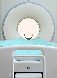
3T MRI
MAGNETOM Verio is the shortest 3T system available today, with an ultra-light magnet. The Magnetom Verio 3T machine is a 70cm open bore MRI system that is equipped with the Tim™ (Total Imaging Matrix) technology that allows for shorter exam times. This system offers more access that will result in more comfort for the patient. This system has been designed for patients of all shapes and sizes. The table on this machine can accommodate patients up to 550 lbs. Patients have less discomfort as well as less anxiety resulting in less sedation The MAGNETOM Verio offers greater patient access and comfort made possible by the 70 cm Open Bore resulting in higher throughput and more referrals. Because patients feel less discomfort and anxiety, it reduces the need for sedation and minimizes claustrophobic rejections. In addition, Tim™ technology also increases throughput thanks to shorter scan time. This machine has a TrueForm™ ultra-light magnet and gradient design that fits the true form of the body. This results in more imaging volume compared to the conventional MRI designs. It also has a higher image quality, better fat saturation as well as the ability to efficiently use the full available field of view of 50×50×45 cm, accessing an anatomical coverage of up to a 50 cm field of view.
- A unique combination of 3T and 70 cm Open Bore
- The shortest 3T system on the market today
- Large field of view, support a full range of clinical applications
- TrueForm™ magnet design offers enhanced image quality by optimizing the homogeneity
Higher speed and superb image quality powered by the VQ-engine gradient.
Pueblo Medical Imaging, a trusted resource for MRI in Las Vegas.
1.5 T AND OPEN MRI


MR imaging uses a powerful magnetic field, radio waves and a computer to produce detailed pictures of organs, soft tissues, bone and virtually all other internal body structures. The images can then be examined on a computer monitor or printed. MRI does not use ionizing radiation (x-rays) and can accomodate up to 660 lbs.
Detailed MR images allow physicians to better evaluate parts of the body for certain diseases that may not be assessed adequately with other imaging methods such as x-ray, ultrasound or computed tomography (also called CT or CAT scanning).
What are some common uses of the MRI procedure?
MR imaging is usually the best choice for examining or evaluating the:
- Body’s major joints
- Spine for disk disease
- Soft tissues of the extremities (muscles and bones)
- Organs of the chest, abdomen and pelvis—including the heart, liver, biliary tract, kidney, spleen, and pancreas and adrenal glands
- Pelvic organs including the reproductive organs in the male (prostate and testicles) and the female (uterus, cervix and ovaries)
- Pelvic and hip bones
- Blood vessels (MR Angiography)
- Breasts
Physicians use the MR examination to help diagnose or monitor treatment for conditions such as:
- Degenerative joint disorders such as arthritis and meniscus tears (knee)
- Fractures (in selected patients)
- Joint abnormalities due to trauma (tendon tears for example)
- Spinal disk abnormalities (herniated disk for example)
- The integrity of the spinal cord after trauma
- Sports-related injuries and work-related disorders caused by repeated strain, vibration or forceful impact
- Tumors (primary tumors and metastases for example) involving bones and joints
- Pain, swelling or bleeding in the tissues in and around the joints and bones
- Tumors of the chest, abdomen or pelvis
- Coronary artery disease and heart problems including the aorta, coronary arteries and blood vessels, by examining the size and thickness of the chambers of the heart and the extent of damage caused by a heart attack or progressive heart disease. For more information, visit the MRA and CT Screening pages
- Tumors and other abnormalities of the reproductive organs (e.g., uterus, ovaries, testicles, prostate)
- Causes of pelvic pain in women, such as endometriosis
- Functional and anatomical abnormalities of the heart
- Diseases of the liver, such as cirrhosis, and that of other abdominal organs (when a complete diagnostic assessment cannot be done with other techniques)
- Congenital arterial and venous vascular anomalies and diseases (e.g., atherosclerosis) of the chest, abdomen and pelvis (MR Angiography)
- Breast cancer and implants
If you are looking for a Las Vegas MRI experience you can trust. Pueblo Medical Imaging is here to provide it.
MRA


In magnetic resonance angiography (MRA), a powerful magnetic field, radio waves and a computer produce the detailed images. MR angiography does not use ionizing radiation (x-rays). MR angiography may be performed with or without contrast material.
What are some common uses of the procedure?
MR angiography is used to examine blood vessels in key areas of the body, including the:
- Brain
- Kidneys
- Pelvis
- Legs
- Lungs
- Heart
- Neck
Physicians use the procedure to:
- Identify disease and aneurysms in the aorta or in other major blood vessels
- Detect atherosclerosis disease in the carotid artery of the neck, which may limit blood flow to the brain and cause a stroke
- Identify a small aneurysm or arteriovenous malformation inside the brain
- Detect atherosclerotic disease that has narrowed the arteries to the legs and help prepare for surgery
- Indicate disease in the renal artery or visualize blood flow to help prepare for a kidney transplant
- Guide surgeons making repairs to diseased blood vessels, such as implanting or evaluating a stent
- Detect injury to one or more arteries in trauma patients
- Evaluate the details of arteries feeding a tumor prior to surgery
- Identify dissection in the aorta or its major branches
- Show the extent and severity of atherosclerosis in the coronary arteries
- Plan for a surgical operation, such as coronary bypass
- Screen individuals for arterial disease, especially patients with a family history of arterial disease or disorders
Trust the Las Vegas professionals at PMI for your MRA services.
CT
 CT scanning—sometimes called CAT scanning—is a noninvasive medical test that helps physicians diagnose and treat medical conditions. CT imaging combines special x-ray equipment with sophisticated computers to produce multiple images or pictures of the inside of the body. These cross-sectional images of the area being studied can then be examined on a computer monitor or printed. CT scans of internal organs, bone, soft tissue and blood vessels provide greater clarity and reveal more details than regular x-ray exams.
 Using specialized equipment and expertise to create and interpret CT scans of the body, radiologists can more easily diagnose problems such as cancers, cardiovascular disease, infectious disease, trauma and musculoskeletal disorders. CT scanning provides more detailed information on head injuries, stroke, brain tumors and other brain diseases than regular radiographs (x-rays). Using CT, the bony structure of the spinal vertebrae is clearly and accurately shown by CT scanning, as are the intervertebral disks and, to some degree, the spinal cord. Perhaps the most frequent use of spinal CT is to detect—or rule out—spinal column damage in patients who have been injured.
What are some common uses of the CT procedure?
CT scanning of the body is:
- One of the best and fastest tools for studying the chest, abdomen and pelvis because it provides detailed, cross-sectional views of all types of tissue.
- Often the preferred method for diagnosing many different cancers, including lung, liver and pancreatic cancer, since the image allows a physician to confirm the presence of a tumor and measure its size, precise location and the extent of the tumor’s involvement with other nearby tissue.
- An examination that plays a significant role in the detection, diagnosis and treatment of vascular diseases that can lead to stroke, kidney failure or even death. CT is commonly used to assess for pulmonary embolism (a blood clot in the lung vessels) as well as for abdominal aortic aneurysms (AAA).
CT scanning of the head is typically used to detect:
- Bleeding, brain injury and skull fractures in patients with head injuries
- Bleeding caused by a ruptured or leaking aneurysm in a patient with a sudden severe headache
- A blood clot or bleeding within the brain shortly after a patient exhibits symptoms of a stroke
- A stroke, especially with a new technique called Perfusion CT
- Brain tumors
CT scanning of the spine is also performed to:
- Evaluate the spine before and after surgery
- Detect various types of tumor in the vertebral column, including those that have spread there from another area of the body. Some tumors that arise elsewhere are first identified by finding deposits of malignant cells (metastases) in the vertebrae; prostate cancer is an example.
- Help diagnose spinal pain. One of the most common causes of spinal pain that may be diagnosed by CT is a herniated intervertebral disk, sometime with CT myelography
- Guide diagnostic procedures such as the biopsy of a suspicious area to detect cancer, or the removal of fluid from a localized infection (abscess)
Physicians often use the CT examination to:
- Quickly identify injuries to the lungs, heart and vessels, liver, spleen, kidneys or other internal organs in cases of trauma
- Guide biopsies and other procedures such as abscess drainages and minimally invasive tumor treatments
- Plan for and assess the results of surgery
- Measure bone mineral density for the detection of osteoporosis
CT scanning is also performed to:
- Evaluate the extent of bone and soft tissue damage in patients with facial trauma, and planning surgical reconstruction
- Diagnose diseases of the temporal bone on the side of the skull, which may be causing hearing problems
- Determine whether inflammation or other changes are present in the paranasal sinuses
- Plan radiation therapy for cancer of the brain or other tissues
- Guide the passage of a needle used to obtain a tissue sample (biopsy) from the brain
- Assess aneurysms or arteriovenous malformations through a technique called CT angiography.
- In patients with narrowing of the spinal canal, vertebral fracture, infection or degenerative disease such as arthritis, CT of the spine may provide important information when performed alone or in addition to magnetic resonance imaging (MRI).
Our Las Vegas CT professionals at Pueblo Medical Imaging (PMI) will ensure your trust.
LOW DOSE CT LUNG CANCER SCREENING
Low dose CT Lung Screening may detect lung cancer before symptoms are being experienced.
This is important as lung cancer has usually metastasized and is often not treatable once symptoms are present.
Low dose CT lung screening is recommended for high-risk individuals with a history of heavy smoking. Have a discussion with your primary care physician about scheduling the exam.
Here are the guidelines put forth by authorities such as American Cancer Society and American Lung Association:
- 55 to 74 years old
- In fairly good health
- Have at least a 30 pack-year smoking history
- Are either still smoking or have quit smoking within the last 15 years
Medicare and many insurances cover this exam. PMI also offers a cash pay option if needed.
CT CORONARY ARTERY SCREENING
 Cardiac CT for Calcium Scoring
A cardiac CT scan is a non-invasive way of obtaining information about the location and extent of calcified plaque in the coronary arteries—the vessels that supply oxygen-containing blood to the heart wall. Plaque is a build-up of fat and other substances, including calcium, which can, over time, narrow the arteries or even close off blood flow to the heart. The result may be painful angina in the chest or a heart attack.
Because calcium is a marker of coronary artery disease, the amount of calcium detected on a cardiac CT scan is a helpful diagnostic tool. The findings on cardiac CT are expressed as a calcium score. Another name for this test is coronary artery calcium scoring.
ULTRASOUND
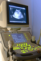 Ultrasound imaging, also called sonography, involves exposing part of the body to high-frequency sound waves to produce pictures of the inside of the body. The ultrasound machine consists of a sophisticated computer connected by a cord to a microphone type device. This device, called a transducer, is actually the combination of a sound transmitter and a microphone. The transducer sends high-pitched sounds into the body. The sounds are above the hearing range for humans. It then listens for the echoes that come back from your tissues. Different tissues of the body send back different echoes. Also, certain diseased or injured tissues send back different echoes than healthy tissues. The computer takes the information from the echoes and turns it into a set of images.
Doppler ultrasound is a special ultrasound technique that evaluates blood as it flows through a blood vessel, including the body’s major arteries and veins in the abdomen, arms, legs, and neck.
 Ultrasound is used to look at soft tissue structures in the abdomen. It can evaluate infection, inflammation, and tumors. It is especially successful in detecting gallstones. Ultrasound is also used to evaluate pelvic structures in women. It is one of the best ways to look for cysts and tumors. Because it uses no radiation, it is also an excellent way of evaluating pregnancies.
PMI is your trusted destination in Las Vegas for Ultrasound services.
- Abdominal Ultrasound
- Vascular Ultrasound
- Obstetrical Ultrasound
- Pelvic Ultrasound
- Renal Ultrasound
- Thyroid Ultrasound
- Testicular Ultrasound
- Pediatric Ultrasound
MAMMOGRAPHY/BONE DENSITY SCREENINGS
Screening Mammogram (2D and 3D)
Diagnostic Mammogram
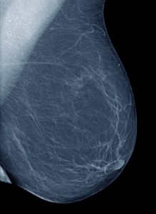
A mammogram is an x-ray examination that uses extremely low doses of radiation to obtain accurate images of the breasts. It is the best way of detecting small cancers even before they can be felt. The American Cancer Society estimates that one in nine women will develop breast cancer in her lifetime. While the exact cause of breast cancer has not been found, we do know that it is treatable and can often be cured when detected early. The American Cancer Society recommends that after consulting with your physician, if you are not experiencing any breast symptoms or problems, you should have a “baseline” mammogram at age 40 and then a yearly screening mammogram. Your physician may recommend a screening mammogram before age forty, based on your risk factors for breast cancer. Because a small percentage of breast cancers cannot be seen on a mammogram, your best defense against breast cancer is an annual mammogram, monthly breast self-examination, and an annual breast exam by your physician.
Bone Densitometry
Osteoporosis is a leading cause of disability among older people, particularly elderly women. Bones weakened by mineral loss are prone to fractures, especially in the spine and hip. Low bone density is measured by a number of techniques. These techniques all measure bone mineralization and detect osteoporosis or bone loss due to menopause, medications or other risk factors. DEXA scan is one of the most widely used detectors of bone mineral density. The use of low dose radiation in conjunction with highly sophisticated computerized analysis provides unique and accurate information about bone density. One of our board-certified radiologists will interpret the results. Your test results will assist your physician in determining if treatment for bone loss is necessary.
MRI OF THE BREAST
Magnetic resonance imaging (MRI) is a noninvasive, usually painless medical test that helps physicians diagnose and treat medical conditions.
MR imaging uses a powerful magnetic field, radio waves and a computer to produce detailed pictures of organs, soft tissues, bone and virtually all other internal body structures. The images can then be examined on a computer monitor or printed. MRI does not use ionizing radiation (x-rays).
Detailed MR images allow physicians to better evaluate parts of the body and certain diseases that may not be assessed adequately with other imaging methods such as x-ray, ultrasound or computed tomography (also called CT or CAT scanning).
MRI of the breast offers valuable information about many breast conditions that cannot be obtained by other imaging modalities, such as mammography or ultrasound. MRI of the breast is not a replacement for mammography or ultrasound imaging but rather a supplemental tool for detecting and staging breast cancer and other breast abnormalities.
Medical studies are currently being conducted to determine whether MRI and other imaging methods can contribute to the early detection and prevention of deaths from breast cancer.
BREAST BIOPSY

Lumps or abnormalities in the breast are often detected by physical examination, mammography, or other imaging studies. However, it is not always possible to tell from these imaging tests whether a growth is benign or cancerous.
A breast biopsy is performed to remove some cells—either surgically or through a less invasive procedure involving a hollow needle—from a suspicious area in the breast and examine them under a microscope to determine a diagnosis. During a breast biopsy, part or all of a tumor may be removed.
Image-guided biopsy is performed when the abnormal area in the breast is too small to be felt, making it difficult to locate the lesion by hand (called palpation).
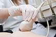
Ultrasound-Guided Biopsy or Aspiration
In ultrasound-guided breast biopsy, ultrasound imaging is used to help guide the interventional radiologist’s instruments to the site of the abnormal growth. There are times when your doctor may decide that ultrasound guidance for biopsy is appropriate even for a mass that can be felt.
Stereotactic Biopsy
In stereotactic breast biopsy, a special mammography machine uses ionizing radiation to help guide the radiologist’s instruments to the site of the abnormal growth. Stereotactic breast biopsy is also performed when the patient or physician strongly prefers a non-surgical method of assessing a breast abnormality.
MRI Breast Biopsy
MRI Breast Biopsy is used to biopsy an abnormal area seen on a Breast MRI in which the abnormal area is either not identified or not seen as clearly on a mammogram or ultrasound. In MRI-guided breast biopsy, magnetic resonance imaging is used to help guide the radiologist’s instruments to the site of the abnormal growth.
FLUOROSCOPY


Studies are performed with a machine called a fluoroscope. This is a device that has a tilting table connected to an x-ray machine and a television screen. The fluoroscope produces a real-time x-ray image on the TV screen. The images on the screen can then be turned into permanent images, which are used for diagnosis. Many fluoroscopic procedures use a liquid contrast. This liquid comes in different forms, which make it much easier for x-rays to show internal parts of the body such as the stomach, esophagus, or colon. Images are taken on the fluoroscope as the contrast material passes through these parts of the body.
Specific studies are listed below:
- Arthrography with Plain x-ray, CT, or MRI
- Barium Enema – Single Contrast, or Air
Contrast, or Gastrograffin
- Barium Swallow
- Upper GI
- Fistulogram
- Hysterosalpingogram
- Routine X-rays or Radiographs
- Small Bowel Follow Through
- Voiding Cystourethrogram
- Pediatric Fluoroscopy
X-RAY
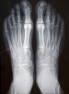
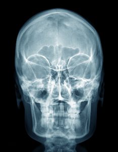
X-rays are a type of electromagnetic radiation used in imaging or therapy that uses short wavelength energy beams which penetrate the body to form an image on film. The x-rays pass through different tissues of the body and onto the film in different amounts. For instance, very few x-rays pass through bones, so they appear white on the film; more pass through muscle, so they appear gray. Metal and contrast media (intravenous or oral contrast) block almost all the photons and will appear bright white. These differences are what create the image on the film. Routine x-rays or plain films are x-rays taken without the use of contrast medium. Many parts of the body, such as chest and bones, are imaged this way. X-rays can help detect pneumonia, fractures, obstructions, tumors, and other diseases.
PMI’s professional staff are trusted leaders for X-Ray in Las Vegas